
What Is KPV?
KPV peptide is a synthetic tripeptide built from three amino acids, lysine, proline, and valine, joined in that sequence (H-Lys-Pro-Val-OH). It corresponds to the C-terminal fragment, residues 11–13, of alpha-melanocyte-stimulating hormone (α-MSH), and is sometimes labeled α-MSH(11–13) or ACTH(11–13) in the older pharmacology literature.
In plain terms, KPV is the smallest piece of the α-MSH hormone that still carries meaningful biological activity in cell and animal studies, but it does so without needing to occupy a melanocortin receptor the way the full hormone does. That distinction is what makes KPV a peptide of interest on its own, separate from α-MSH or its receptor-targeting analogs.
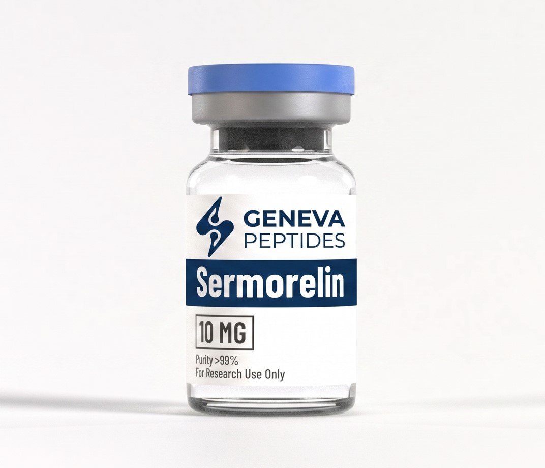
Geneva Peptide supplies KPV as a lyophilized powder for laboratory research use only. It is manufactured and sold strictly for use by qualified researchers in controlled settings, not as a drug, supplement, cosmetic ingredient, or article for human or animal use.
What’s Included?
| Detail | Specification |
|---|---|
| Vial & net content | 10 mg of KPV peptide per vial, weighed and sealed under inert atmosphere. |
| Physical form | Lyophilized (freeze-dried), white to off-white powder or friable cake. |
| Purity | ≥99% by reverse-phase HPLC, confirmed against a reference standard. |
| Documentation | Batch-specific Certificate of Analysis (COA); SDS available on request. |
KPV Technical Specifications
Every physical and chemical constant below describes the KPV peptide molecule itself, independent of batch, batch-level purity and identity data live on the Certificate of Analysis for each lot.
| Specification | Details |
|---|---|
| Sequence | H-Lys-Pro-Val-OH |
| Also known as | α-MSH (11–13); ACTH (11–13); Msh(11-13) |
| Molecular formula | C₁₆H₃₀N₄O₄ |
| Molecular weight | 342.4 g/mol |
| CAS number | 67727-97-3 |
| PubChem CID | 125672 |
| Purity (this lot) | ≥99% (HPLC) |
| SKU | GP-KPV-10 |
How KPV works?
Unlike full-length α-MSH, KPV does not require binding to a melanocortin receptor (MC1R–MC5R) to exert measurable effects in cell models. Research to date points to three overlapping, receptor-independent mechanisms:
| Mechanism | Description |
|---|---|
| NF-κB nuclear translocation blockade | KPV has been reported to compete with importin-α for binding to the p65/RelA subunit of NF-κB at its armadillo-repeat domains, which keeps the transcription factor trapped in the cytoplasm and prevents it from switching on pro-inflammatory gene expression in the nucleus (Kelly et al., 2006). |
| MAPK pathway modulation | In stressed epithelial and keratinocyte models, KPV has been associated with reduced phosphorylation of p38 and JNK, two MAPK-family kinases that normally amplify inflammatory and oxidative-stress signaling. |
| PepT1-mediated cellular uptake | As a tripeptide, KPV is a substrate for PepT1 (SLC15A1), the oligopeptide transporter expressed on intestinal epithelium and select immune tissues. This transporter appears to be the route by which KPV reaches its intracellular targets in gut-inflammation models, and its effect is substantially reduced in PepT1 knockout animals. |
Research Applications & Scientific Background
Published research on KPV clusters around four areas. All findings below come from in vitro or animal studies; KPV has not completed controlled human clinical trials for any indication.
Intestinal inflammation models
In DSS- and TNBS-induced colitis models, KPV administration has been associated with reduced weight loss and lower histologic inflammatory infiltrate, an effect that depends on PepT1-mediated uptake and is diminished in PepT1-knockout mice. Related work has also examined KPV in colitis-associated cancer models.
Epithelial & keratinocyte signaling
In human keratinocyte cultures, KPV has been shown to suppress IL-8 production at concentrations as low as 10 nM without engaging MC1R or raising intracellular cAMP. More recent work has extended this to fine-particulate-induced oxidative stress, where KPV modulated MAPK/NF-κB signaling in keratinocyte models.
Systemic inflammation & endotoxemia models
A dimerized form of the peptide, (CKPV)₂, reduced mortality from roughly 80% to 10% in a mouse endotoxemia model at 4 mg/kg, a result frequently cited as evidence for KPV-class peptides acting on systemic, not just local, inflammatory responses.
Antimicrobial activity (in vitro)
Separate in vitro screens have reported antimicrobial activity for KPV against organisms including Staphylococcus aureus and Candida albicans, though this remains an early and comparatively small body of evidence.
KPV vs. Related Compounds
KPV is frequently discussed alongside its parent hormone and a handful of structurally or functionally adjacent peptides. The table below is a research-orientation aid, not an equivalence claim, each compound has a distinct mechanism and evidence base.
| Compound | Structure / class | Receptor dependence | Primary research focus |
|---|---|---|---|
| KPV | Lys-Pro-Val tripeptide, α-MSH(11–13) | Receptor-independent (NF-κB / MAPK) | Mucosal & epithelial inflammation |
| α-MSH (full length) | 13-residue native melanocortin hormone | MC1R / MC3R / MC4R-dependent | Pigmentation, appetite, systemic melanocortin signaling |
| KdPT | Lys-D-Pro-Thr, α-MSH-derived analog | Receptor-independent | Proteolytically stabilized variant of the KPV pharmacophore |
| Ac-KPV-NH₂ | Acetylated, amidated KPV analog | Receptor-independent | Extended-half-life formulation research |
| BPC-157 | Partial sequence, gastric-derived pentadecapeptide | Not fully characterized | Tissue repair & angiogenesis research (distinct pathway) |
Scientific References
- 01 Kelly, C.J. et al. (2006). Characterization of the mechanism by which the α-MSH(11–13) tripeptide KPV inhibits NF-κB nuclear translocation via competition with importin-α for p65/RelA binding.
- 02 Elliott, R.J. et al. (2004). Suppression of IL-8 secretion in human keratinocytes by the α-MSH fragment KPV, independent of MC1R engagement.
- 03 Dalmasso, G. et al. (2008). Gastroenterology — PepT1-mediated uptake of the tripeptide KPV reduces intestinal inflammation in murine colitis models.
- 04 Gatti, S. et al. (2006). Protective effect of the (CKPV)₂ dimer against lethal endotoxemia in a murine sepsis model.
- 05 Cells (2024–2025). KPV modulation of MAPK/NF-κB signaling in keratinocytes challenged with fine particulate matter.
Reference list is provided for research orientation. Verify full citations (journal, volume, DOI) against primary literature before external publication.
Storage & Laboratory Handling
Storage conditions
Store lyophilized KPV tightly sealed at −20°C, protected from light and moisture, ideally with desiccant. Under these conditions the unreconstituted peptide is expected to remain stable for approximately 2–3 years. For short-term storage, 2–8°C is acceptable for up to several months. Avoid repeated freeze–thaw cycles of any solution.
Laboratory handling
Handle KPV only in a well-ventilated area, ideally under a fume hood or laminar-flow enclosure, wearing standard PPE (gloves, eye protection, lab coat). Avoid contact with skin, eyes, or clothing, and avoid inhalation or ingestion. KPV is not classified as hazardous under OSHA HazCom 2012, but its toxicological profile has not been fully characterized — treat it as a substance of unknown toxicity.
Reconstitution notes for research use
When reconstituting for in vitro or in vivo research protocols, add diluent slowly down the vial wall and swirl gently rather than vortexing, to protect peptide structure. Reconstituted solution stability is diluent- and condition-dependent; researchers commonly report usable stability of roughly 5–14 days when stored at 2–8°C and protected from light. Aliquot and freeze any solution not used promptly to avoid freeze–thaw degradation of the working stock.
Frequently Asked Questions
What is KPV peptide used for in research?
In laboratory settings, KPV is studied primarily as a tool compound for probing melanocortin-receptor-independent inflammatory signaling, particularly NF-κB and MAPK pathway activity in intestinal, epithelial, and immune-cell models.
How is KPV different from full alpha-MSH?
Full-length α-MSH is a 13-residue hormone that acts by binding melanocortin receptors (MC1R–MC5R), producing pigmentary and appetite-related effects. KPV is just the C-terminal three residues and appears to act independently of those receptors, which is why researchers treat it as a distinct compound rather than a simple α-MSH fragment.
Does KPV require melanocortin receptor binding to be active?
No. Published cell studies report KPV activity, such as IL-8 suppression in keratinocytes, at concentrations where MC1R is not engaged and intracellular cAMP does not rise, supporting a receptor-independent mechanism.
What purity does Geneva Peptide’s KPV meet?
Each 10 mg vial is manufactured to ≥99% purity, confirmed by reverse-phase HPLC and mass spectrometry, with lot-specific results provided on the Certificate of Analysis.
How should KPV be stored before reconstitution?
Keep the lyophilized powder sealed at −20°C, protected from light and moisture. Short-term storage at 2–8°C is acceptable for up to several months.
How long is reconstituted KPV stable?
Stability depends on diluent and storage conditions; researchers commonly report roughly 5–14 days at 2–8°C. Aliquoting and freezing unused solution helps avoid degradation from repeated freeze–thaw cycles.
Is KPV approved for human or veterinary use?
No. KPV is sold strictly for laboratory research use by qualified researchers. It is not approved by the FDA or any equivalent body for human or veterinary use, and Geneva Peptide does not sell it for those purposes.
What documentation ships with each vial?
Every order includes a batch-specific Certificate of Analysis covering identity and purity testing. A Safety Data Sheet (SDS) is available on request.